Home
/ Coronal View Of Brain, brain anatomy | MRI coronal brain anatomy | free MRI cross ... - A coronal section is one that separates the brain into anterior and posterior halves.
Coronal View Of Brain, brain anatomy | MRI coronal brain anatomy | free MRI cross ... - A coronal section is one that separates the brain into anterior and posterior halves.
Coronal View Of Brain, brain anatomy | MRI coronal brain anatomy | free MRI cross ... - A coronal section is one that separates the brain into anterior and posterior halves.. Hd00:33an abnormal mri scan of the brain depicting demylenating lesions often associated with multiple sclerois. Aka frontal plane, a coronal plane divides the brain into an anterior and posterior portion. Whenever necessary, information from the extensive literature pertaining to the atlas brain was integrated. Medial view of the right cerebrum. Brain, coronal section, basal ganglia, close view, gross.
I also got some of the coloring books for neuroanatomy, but alas. Viewing the circle of willis as an angry spider). A coronal section is one that separates the brain into anterior and posterior halves. The frontal and temporal lobes are observed in their previously described locations. Learn vocabulary, terms and more with flashcards, games and other study tools.
Brain Anatomy Jeopardy Template from i.ytimg.com It is one of the three main planes of the body used to describe the location of body parts in relation to each other axis. Coronal cut of the human brain in which we can see the brain composed of two halves, one right and one left, in this illustration. The diencephalon is represented by a posterior view of the thalamus with its nuclei, followed by a sagittal section to locate the hypothalamus (including mammillary bodies, tuber cinereum, lamina terminalis), the epithalamus (with the pineal gland and. (a) coronal view through the frontal horns shows multiple calcifications in the region of the thalami and basal. Coronal t2 weighted magnetic resonance image of the brain view full text. Transverse (coronal) sections rostral to caudal. Whenever necessary, information from the extensive literature pertaining to the atlas brain was integrated. A coronal section of the head is viewed and interpreted from the point of view that the clinician is facing the patient.
Normal sagittal and coronal views of the infant brain.
A coronal plane (also known as the frontal plane) is any vertical plane that divides the body into ventral and dorsal (belly and back) sections. (a) coronal view through the frontal horns shows multiple calcifications in the region of the thalami and basal. Free online quiz coronal view of the brain 1. Anatomy of the larynx 3d medical illustration on white background. Whenever necessary, information from the extensive literature pertaining to the atlas brain was integrated. Coronal cut of the human brain in which we can see the brain composed of two halves, one right and one left, in this illustration. A coronal section of the head is viewed and interpreted from the point of view that the clinician is facing the patient. It is created by slicing the brain parallel to the. The review of the literature revealed that optic neuritis represents the 46.4% (124/267) of cranial neuritis in ss patients [7, 18, 19, 24, 34, 36 in a few cases, neuritis was associated with cns involvement, such as brain vasculitis 34,77, acute transverse myelopathy 43,67, myelitis [77,80,83. Cta brain or computed tomographic angiography of the brain coronal view / 3d rendering image showing vessels in human brain. Home › create › flashcards › health › human body › brain › coronal brain. Professor sarah meerts highlights different sections of a dissected human brain. It is one of the three main planes of the body used to describe the location of body parts in relation to each other axis.
The review of the literature revealed that optic neuritis represents the 46.4% (124/267) of cranial neuritis in ss patients [7, 18, 19, 24, 34, 36 in a few cases, neuritis was associated with cns involvement, such as brain vasculitis 34,77, acute transverse myelopathy 43,67, myelitis [77,80,83. Whenever necessary, information from the extensive literature pertaining to the atlas brain was integrated. The frontal and temporal lobes are observed in their previously described locations. Illustration of coronal view of a human brain, including the corpus callosum, pons, hippocampus and caudate nucleus. Hippocampus , medial view/coronal section.
100 MICRON MRI OF THE HUMAN BRAIN - CORONAL - YouTube from i.ytimg.com Anatomy of the larynx 3d medical illustration on white background. The coronal section shown in figure 19a occurs approximately halfway between these two poles, and the ventral view of figure 19b identifies several major structures that appear on the bottom surface of the brain. Whenever necessary, information from the extensive literature pertaining to the atlas brain was integrated. My brain doctor recommended clinical neuroanatomy made ridiculously simple, which i find to be quite useful as there are a lot of analogies and metaphors integrated into the text (i.e. Transverse (coronal) sections rostral to caudal. Normal sagittal and coronal views of the infant brain. A coronal section of the head is viewed and interpreted from the point of view that the clinician is facing the patient. Professor sarah meerts highlights different sections of a dissected human brain.
Coronal view of the brain and its features in mri section for more information visit website akaarstar.wordpress.com for the kshtrgyn website visit kshtrgyn.
I also got some of the coloring books for neuroanatomy, but alas. Normal sagittal and coronal views of the infant brain. Upload, livestream, and create your own videos, all in hd. Examples in the 3d reconstruction section show different views of the thalamus and some additional landmark structures. Professor sarah meerts highlights different sections of a dissected human brain. Free online quiz coronal view of the brain 1. The review of the literature revealed that optic neuritis represents the 46.4% (124/267) of cranial neuritis in ss patients [7, 18, 19, 24, 34, 36 in a few cases, neuritis was associated with cns involvement, such as brain vasculitis 34,77, acute transverse myelopathy 43,67, myelitis [77,80,83. Home › create › flashcards › health › human body › brain › coronal brain. The frontal and temporal lobes are observed in their previously described locations. Professor sarah meerts highlights different sections of a dissected human brain. Learn vocabulary, terms and more with flashcards, games and other study tools. Hd00:33an abnormal mri scan of the brain depicting demylenating lesions often associated with multiple sclerois. 0 ratings0% found this document useful (0 votes).
Hippocampus , medial view/coronal section. Coronal t2 weighted magnetic resonance image of the brain view full text. The frontal and temporal lobes are observed in their previously described locations. Home › create › flashcards › health › human body › brain › coronal brain. The coronal section shown in figure 19a occurs approximately halfway between these two poles, and the ventral view of figure 19b identifies several major structures that appear on the bottom surface of the brain.
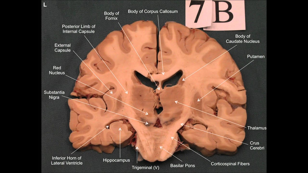
2: Coronal slices of human brain. A coronal slice of an ... from www.researchgate.net Therefore, the patient's left side will be on coronal section of the brain at the level of the thalamus. Case contributed by dr maciej debowski. Illustration of coronal view of a human brain, including the corpus callosum, pons, hippocampus and caudate nucleus. The coronal section shown in figure 19a occurs approximately halfway between these two poles, and the ventral view of figure 19b identifies several major structures that appear on the bottom surface of the brain. 0 ratings0% found this document useful (0 votes). The frontal and temporal lobes are observed in their previously described locations. Free online quiz coronal view of the brain 1. Medial view of the right cerebrum.
Anatomy of the larynx 3d medical illustration on white background.
Transverse (coronal) sections rostral to caudal. Examples in the 3d reconstruction section show different views of the thalamus and some additional landmark structures. Learn vocabulary, terms and more with flashcards, games and other study tools. Coronal t2 weighted magnetic resonance image of the brain view full text. Viewing the circle of willis as an angry spider). Hippocampus is visible after removing brain stem and most of the right thalamus. (a) coronal view through the frontal horns shows multiple calcifications in the region of the thalami and basal. My brain doctor recommended clinical neuroanatomy made ridiculously simple, which i find to be quite useful as there are a lot of analogies and metaphors integrated into the text (i.e. Coronal cut of the human brain in which we can see the brain composed of two halves, one right and one left, in this illustration. Case contributed by dr maciej debowski. A coronal section of the head is viewed and interpreted from the point of view that the clinician is facing the patient. Illustration of coronal view of a human brain, including the corpus callosum, pons, hippocampus and caudate nucleus. Anatomy of the larynx 3d medical illustration on white background.